DeteCT Vet | Advanced Veterinary CBCT 3D Imaging System

Cut costs, save space, and capture every detail

Transform your practice with DeteCT Vet™, the high-resolution 3D Cone Beam CT (CBCT) system engineered for fast, in-clinic diagnostics. Get full 3D anatomical scans in under 20 seconds using standard wall power, while eliminating the massive footprint, radiation, and facility costs of traditional CT systems. Schedule your demo today to bring advanced pre-surgical planning in-house.

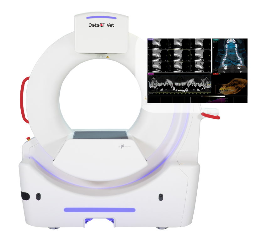
Veterinarian holding a dog next to the DeteCT Vet 3D CBCT scanner showing high-resolution chest and skeletal scan previews
Speed, Precision, Space Savings, and Unbeatable Support.

All in one powerful system

Speed Without Sacrifice

Get a complete 3D scan in under 20 seconds.

Clarity You Can Trust

Large field of view & sharp, detailed images.

Space-Saving Design

Perfect for clinics where every square foot counts.

Unmatched Support

Training, remote help, and the peace of mind of JPI’s warranty.

Switching Between Imaging Modalities in Just One Click Away

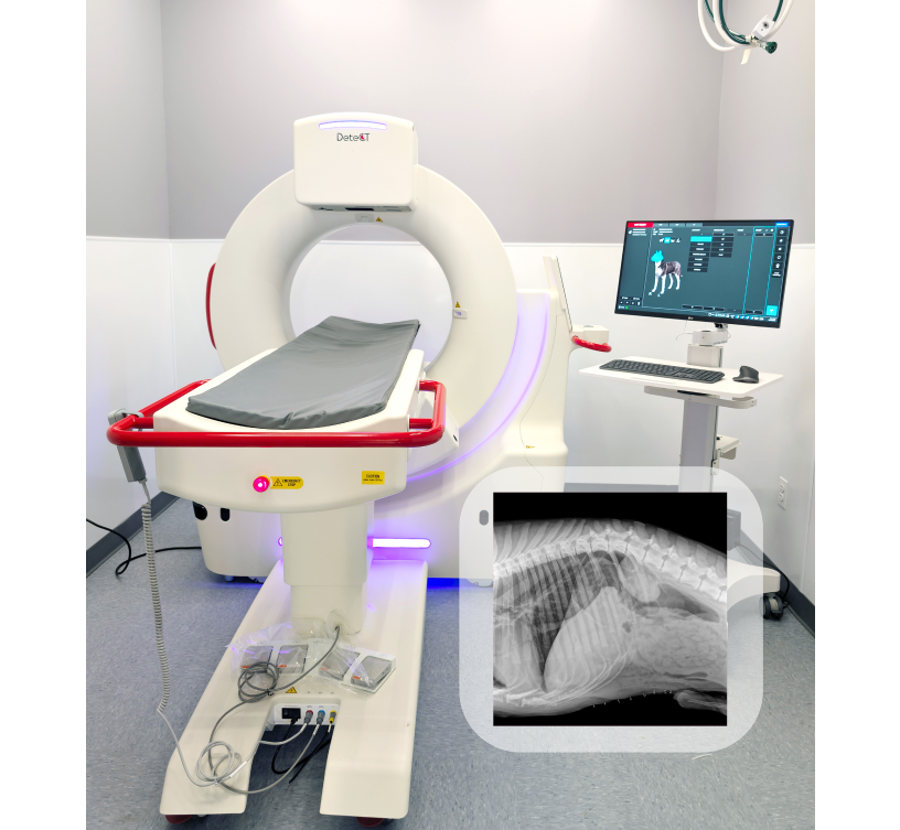
This system lets you switch between CT, radiography, dental panoramic, thermal imaging, and fluoroscopy with just one click—so you can move faster, work smarter, and get to answers sooner

DeteCT Vet 3D CBCT system with patient table, Golden Retriever, and sample 3D anatomical, fluoroscopy, and X-ray scan previews
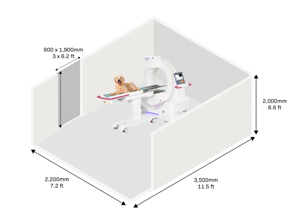
3D isometric diagram of a room layout displaying the DeteCT Vet scanner with a golden retriever lying on the scanning bed.

Hardware, Physical & Operational Specifications

Bore Diameter: 29.5 inches large-bore gantry (crucial for positioning various small animal breed sizes).

Active Field of View (FOV): 17 x 17 inches full flat-panel active imaging area.

Dimensions: Width: 5.7 ft, Height: 5.8 ft.

Mobility & Structural Requirements: Wheeled unit with 360-degree slip-ring rotation; fits in standard clinical rooms as small as 8 x 12 ft.

Power Input: 220V 15A Single Phase

Radiation & Shielding: Low-dose Cone Beam CT (CBCT) architecture; drastically reduces room lead-shielding costs compared to conventional fan-beam CT.

Scan Speed: Scan time acquisition 19s + 46s reconstruction

Frequently Asked Questions

What is the difference between traditional veterinary CT and Cone Beam CT (CBCT)?

Traditional CT uses a fan-shaped X-ray beam and multiple detector rows to create detailed cross-sectional images, while Cone Beam CT (CBCT) uses a cone-shaped beam and flat-panel detector to capture high-resolution 3D images in a compact system. JPI Animal Health’s DeteCT Vet uses CBCT technology to bring fast, high-resolution 3D imaging into veterinary practices with a smaller footprint and lower infrastructure requirements than many conventional CT systems.

Can veterinarians perform 3D imaging in-house?

Yes. Compact veterinary CBCT systems make it possible for veterinary practices to perform high-resolution 3D imaging in-house instead of referring every advanced imaging case to another facility. JPI Animal Health’s DeteCT Vet provides 3D CBCT imaging for detailed anatomical evaluation and pre-surgical planning in a compact system designed for veterinary environments.

What software powers JPI Animal Health imaging systems?

JPI Animal Health imaging systems are powered by ExamVue™ Duo, JPI’s imaging software platform for veterinary X-ray and advanced imaging workflows. ExamVue Duo supports image acquisition, processing, viewing, and DICOM integration while providing a user-friendly workflow designed for veterinary practices.

We’re here to help you

Let's talk about diagnostics solutions